- Joined
- Oct 7, 2008
- Messages
- 61,459
- Location
- Bulgaria

Daniela Poggiali currently 53 years old but was just 42 in 2014 when she murdered 38 of her hospital patients and took photos with the dead still in their hospital beds where she killed them with lethal injections containing various substances in The Umberto I hospital in Lugo in northern Italy.
She was sentenced to 30 years in prison.
Here we present an autopsy case of one of her victims a 77-year-old male who was admitted to the hospital after a stroke.
After just 8 days the patient died of cardiac arrest with very severe respiratory acidosis and hypoxemia.
An autopsy was ordered. The investigators managed to prove her guilt without doubt by obtaining video surveillance from cameras at a local supermarket not far from the hospital where she bought hydrochloric acid which she sprayed directly into the mouth of the elderly patient with a 60ml syrine.
The victim was the father of her colleague and she wanted to take revenge against the patient’s daughter for managing to stay at the hospital while she herself was getting transferred to another.
Meanwhile, the forensic police laboratories identified the caustic substance present in the patient’s clothes as hydrochloric acid - an acid identical to that purchased by the nurse days earlier.
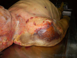
At autopsy:
There were numerous chemical burn injuries affecting the head, the neck, and the right shoulder with varying degrees of severity. These were erythematous areas at times with complete destruction of the surface lining of the epidermis. The shoulder injury was vast and severe. In fact, in this context the chemical burn was so extensive with superficial necrosis of the epidermis that the epidermal vascular reticulum was clearly evident.
In the posterior region of the neck, there was another lesion of the epidermis that was likely caused by posterior drainage of the chemical.
The mouth was affected by numerous erythematous lesions, erosions, and even ulcerations of the skin and/or superficial mucous lining. The palate was also covered with yellowish necrotic material.
By carefully examining the main injured organs, the whole tongue was covered with a yellowish patina of superficial and easily desquamating necrotic material; vascular reticulum was easily observed and lateral to the tongue small erosions/ulcers were evident.
Below the vocal cords a roundish blackish-gray lesion was clearly evident; this finding could be interpreted as the point of contact of the jet of substance with the upper respiratory tract. In the proximal third of the esophagus there was a blackish gray lesion similar to the laryngeal one but less extensive see.
The stomach was significantly altered due to the presence of two ulcerated but not perforated and bleeding areas—one very extended from the cardia to the gastric fundus and a smaller one in the antral site. These viscera contained abundant brownish blood material.
There was no perforation of the airways but only superficial necrosis and very strong irritation. The right lung appeared completely subverted in its structure and altered by the presence of solid, nodular, white-yellowish areas. The left lung was affected by similar phenomena but to a lesser extent see. There was a modest bilateral serum-hematic pleural effusion.
With the examination of the brain, an area of corticalsubcortical necrosis of 10x4 cm, compatible with the previous stroke known in the anamnesis, at the temporoparietal site, was highlighted in the right cerebral hemisphere.
On histological examination, the tongue, pharynx, larynx, and trachea showed complete necrosis of the superficial epithelium with involvement of the subepithelial connective tissue. In the tongue, necrosis also involved the muscular structures of this organ at various locations. In the trachea, cartilage structures were sometimes involved. The lungs were affected by acute bronchopneumonia with diffuse parenchymal necrosis.
The esophagus and stomach showed massive necrosis of the superficial epithelium extending up to the muscular layer. Since a few days had passed between the chemical insult and the patient’s death, the phenomena of necrosis caused by the acid were associated with extensive acute inflammation in all the areas concerned. Therefore, overall, the histological findings were characterized by extensive tissue necrosis associated with acute inflammation In the central nervous system, there was extensive cerebral necrosis associated with gliosis, mainly cortical but also in the surrounding structures in correspondence with the area affected by the earlier ischemic stroke, which had previously been described macroscopically.
The findings collected were compatible with the forced ingestion of a caustic substance caused by another person. In fact, the right lung was massively affected by the lesions.
This was likely because the right lung is connected to the trachea by the right bronchus, which has a more rectilinear course than the contralateral. The left bronchus has a more curved course and this anatomical detail preserved the left lung from more significant involvement. In addition, the upper digestive system together with the upper respiratory tract were simultaneously affected by injuries.